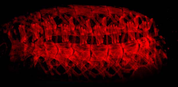
Muscle pattern of a finally developed embryo
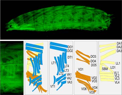
Myogenesis is an evolutionary conserved developmental process characterized by distinct cellular events such as muscle founder determination, cell fusion, myotube extension and anchorage. The development of the body wall musculature of the Drosophila larvae serves as a simple model system for studying conserved aspects of myogenesis and muscle pattern formation during animal embryogenesis. In Drosophila, the musculature consists of about 30 segmentally repeated, stereotypically arranged muscles, which are positioned into three layers. The musculature differentiates during embroygenesis and is used for the active hatching of the finally developed embryo. These muscle are used dring all three larvae stages, they just growth up to 50fold in size, however no new cells are inluded or any division occurs.

Muscle pattern of a larvae and a schematic view of the three muscle layer
Each muscle fibre originates from a distinct muscle founder cell that is determined by specific combinations of transcription factors. Subsequently, the founder cells form syncitial myotubes that extend by the formation of polar growth cone-like structures while fusing with competent myoblasts. As a result, myotubes elongate and extend along the epidermis towards their apodemes at the segment borders.

Each muscle originate from a single muscle founder (left), fuses with myoblasts (center) and grow towards its later attachment sites within the epidermis (right)
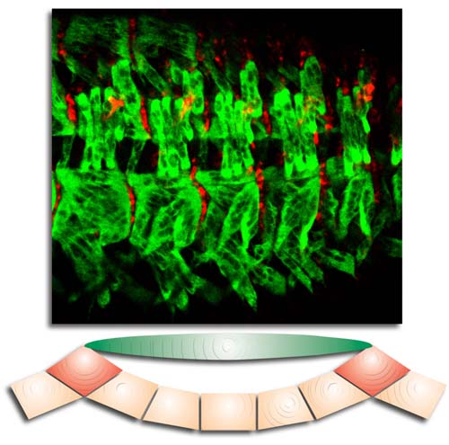
Mutant studies suggest, that signalling processes between the prospective apodemes and the extending myotube regulate directed extension of the myotubes. After contacting each other, myotubes and apodemes start their final differentiation, a process characterized by the expression of proteins of the contractile apparatus. The anchorage of the muscles is assured by integrins that interact with extracellular matrix proteins including Thrombospondin. So both directed migration and functional anchorage of the somatic muscles are controlled only by the direct interaction of differentiating muscles and epidermal muscle attachment cells, the apodemes.

Each muscle is attached to epidermal attachment sites


